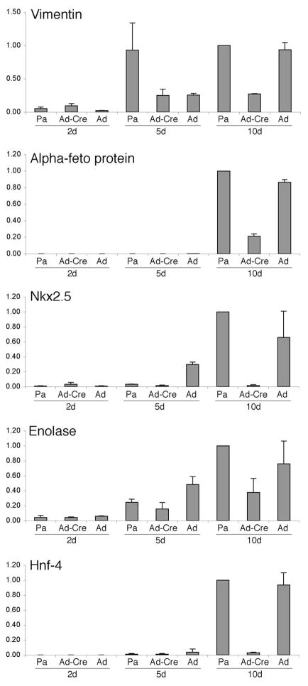
Fig. 5. Expression of Lineage markers in Embryoid Bodies.
Real time RT-PCR analysis of genes expressed in differentiating embryoid bodies over time. Markers for all three germ layers were used, including ectoderm (Vimentin and Enolase), endoderm (β-feto protein and Hnf4), and mesoderm (Nkx2.5). Parental ES ptipfl/− cells (Pa), Ad-Cre 1 infected cells (Ad-Cre), and adenoviral vector infected cells (Ad) were used to generate embryoid bodies in hanging drops then cultured for 2, 5, or 10 days in Petrie dishes. Total RNA was isolated and RT-PCR were normalized to GAPDH. Averages for three readings are shown; error bars represent one standard deviation from the mean.

