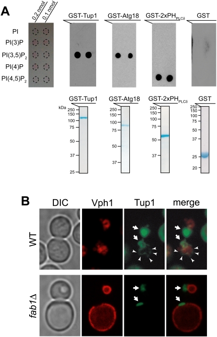
Figure 1.
Tup1 binds PI(3,5)P2 lipid with a high specificity. (A, top panel) GST-Tup1 and GST-Atg18 bound PI(3,5)P2 with a high specificity, while GST-PHPLCδ specifically bound PI(4,5)P2. GST protein alone did not bind any PI lipid. (Bottom panel) Equivalent amount of purified, bacterially expressed GST fusion proteins (50 μg) were used for protein–lipid overlay assay. (B) Vph1-mCherry marks the limiting membrane of vacuoles (red). A pool of GFP-Tup1 localized at the vacuolar membrane (arrowhead), in addition to the nuclear Tup1 (arrow), was observed in some wild-type (WT) cells (see Supplemental Fig. S2B). In contrast, the vacuolar localization of Tup1 was not observed in fab1Δ cells.

