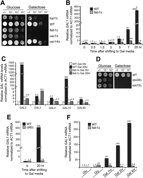
Figure 2.
PI(3,5)P2 and Cti6 are required for transcriptional induction of GAL1. (A) SEY6210 fab1Δ and vac7Δ cells exhibited the Gal− phenotype. SEY6210 vac14Δ cells exhibited the Gal+ phenotype. SEY6210 tup1Δ cells showed slower Gal growth than wild-type (WT) cells. (B) RT-qPCR analysis showed gradual induction of GAL1 mRNA in SEY6210 wild-type cells. At 20 h after Gal shift, a 490-fold induction of GAL1 mRNA was observed in wild-type cells. No detectable increase in GAL1 mRNA was observed in fab1Δ cells at 20 h after Gal shift. The results that are shown with a standard deviation are from three independent analyses. (C) The mRNA levels of GAL2 and GAL10 were highly induced in SEY6210 wild-type cells, but remained constitutively repressed in SEY6210 fab1Δ cells at 20 h after Gal shift. The mRNA levels of GAL3, GAL4, and GAL10 were induced a few-fold in SEY6210 wild-type cells, while, in fab1Δ cells, the levels of GAL4 and GAL80 mRNA remained repressed (see a caveat in the text for the GAL3 mRNA results of fab1Δ cells). (D) SEY6210 vps34Δ and vps15Δ cells, which produce no PI(3)P and PI(3,5)P2, exhibited the Gal− phenotype. (E) SEY6210 cti6Δ cells exhibited a severe defect in GAL1 mRNA induction. (F) SEY6210 cells grown in Glu medium were shifted to Gly medium and grown for several hours to derepress GAL genes. When shifted to Gal medium, GAL1 mRNA was induced very rapidly in wild-type cells, whereas GAL1 remained constitutively repressed in fab1Δ cells.

