Figure 3.
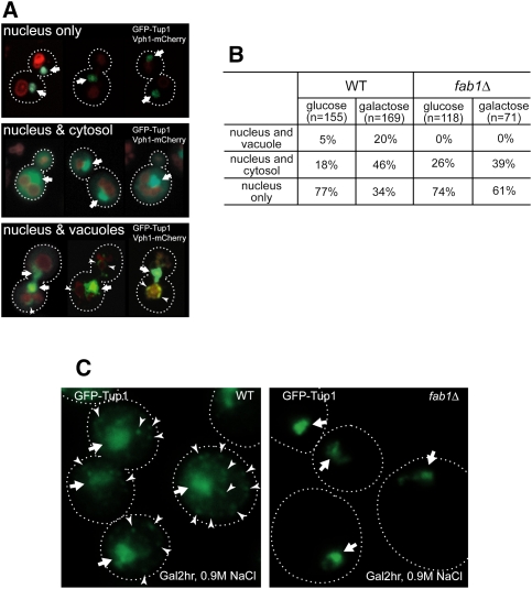
Gal and hyperosmotic stress redistribute Tup1. (A) Montages of cells containing nuclear GFP-Tup1 only (top panel), nuclear and cytosolic GFP-Tup1 (middle panel), or nuclear and vacuolar GFP-Tup1 (bottom panel) and Vph1-mCherry. Nuclear GFP-Tup1 is indicated by an arrow and vacuolar membrane GFP-Tup1 is marked by an arrowhead. (B) Cells grown in Glu or Gal (2 h after shift) medium were imaged, analyzed, and scored for one of the three categories of GFP-Tup1 localization (nuclear only, nuclear and cytosolic, or nuclear and vacuolar membrane). Gal medium significantly enhanced cytosolic and vacuolar membrane localization of Tup1 in wild-type (WT) cells. In contrast, although Gal medium increased cytosolic localization of Tup1 in fab1Δ cells, vacuolar membrane localization of Tup1 was not observed. (C) When wild-type cells in Gal medium were treated with 0.9 M NaCl, Tup1 formed puncta (arrowhead) in the cytoplasm in most of the wild-type cells in addition to the nuclear Tup1 (arrow), whereas Tup1 did not form cytoplasmic puncta in fab1Δ cells.

