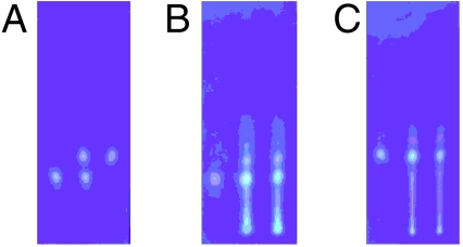
Fig. 11.
TLC detection of mycolactones A/B and C. Photograph A: synthetic mycolactones A/B (left), C (right), and their mixture (center). Photograph B: synthetic mycolactone A/B (left), a lipid extract of an African strain of M. Ulcerans (right), and their mixture (center). Photograph C: synthetic mycolactone C (left), a lipid extract of an Australian strain of M. Ulcerans (right), and their mixture (center).

