Fig. 4.
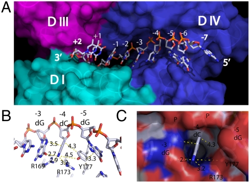
Structural determinants for substrate binding and specificity. (A) Surface representation of the DNA binding groove. The cleavage site sits on the border between domain III and domain IV. (B) The phenol side chain of Y177 wedges between bases -4 and -5 and forms π-interactions among them. (C) The cavity formed at the -4 position can accommodate only a cytosine.

