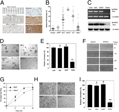
Fig. 1.
Srx is highly expressed in patients with lung cancer and is required for anchorage-independent colony formation, migration, and invasion of cancer cells. (A) Screenshots of representative tissue microarray images (Left) and microscopic images of Srx expression in normal, squamous cell carcinoma (SCC) and adenocarcinoma (AC) (Right). (Scale bar, 10 μm.) (B) Quantitative results of A. NTD, nontumor diseases; mSCC, metastatic SCC. Data are presented as means ± SD. Compared with normal, #P > 0.05, *P < 0.05, **P < 0.01 (t test). See also Table S1 for detailed tissue information. (C) Knockdown of endogenous Srx indicated at the transcript level by RT-PCR and at the protein level by Western blot. (D–I) Srx knockdown cells form fewer colonies in anchorage-independent growth in soft agar (D and E), migrate slower in the wound-healing assay (F and G), and are less invasive in the matrigel invasion assay (H and I). Representative images from sextuplicates are shown. (D, F, and H) Colonies with diameter >100 μm (scale bar in D) were counted. All data are presented as means ± SD (n = 6). (E, G, and I) Compared with the control parental cells, #P > 0.05 and **P < 0.01 (t test in E and I, paired t test in G).

