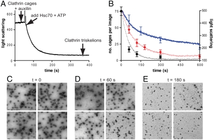
Fig. 1.
A real-time in vitro assay for clathrin cage disassembly and correlation with electron microscopy images of clathrin cages. (A) Representative trace of the right-angle light-scattering assay for clathrin cage disassembly. Clathrin cages (0.09 μM triskelia) were premixed with 0.1 μM auxilin, and after 60 s, cage disassembly was initiated by addition of 1 μM Hsc70 and 500 μM ATP. (B) Average results for three different disassembly assays monitored both by light scattering as in A and compared with electron microscopy images as in C–E. The single points represent the average number of cages counted per image, initiated with 0.1 μM (triangles), 0.2 μM (circles), or 0.5 μM (squares) Hsc70. Data are mean ± SD. The single lines represent the light-scattering results obtained under the same conditions. (C–E) Representative transmission electron micrographs of negatively stained grids prepared at 0, 60, and 180 s during a disassembly assay containing clathrin cages (0.09 μM triskelia), 0.1 μM auxilin, 500 μM ATP, and initiated with 0.2 μM Hsc70. The scale bar in the bottom left-hand corner of each image represents 0.2 μm.

