Fig. 2.
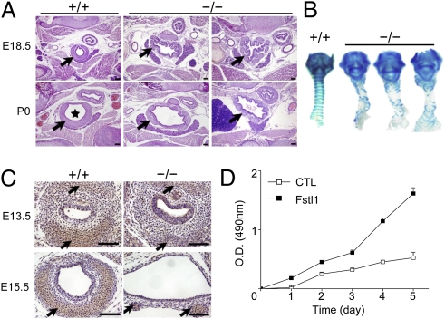
Tracheal malformation in Fstl1−/− mice. (A) Transverse sections of the middle portion of trachea from a WT and two homozygous mice before (E18.5) and after breath (P0) showed an increase in the lumen diameter (asterisk), dispersion and discontinuity of cartilage ring (arrow), and disorganization of the epithelial layer in the mutants. (Scale bars, 100 μm.) (B) Alcian blue staining revealed impaired banding pattern of tracheal C-ring cartilage in all mutant skeletal preparations (ventral views). (C). Sections stained with an antibody against type II collagen (arrows). (Scale bars, 100 μm.) (D). Effects of stable overexpression of Fstl1 on cell proliferation of ATDC5 cells with MTT assays.

