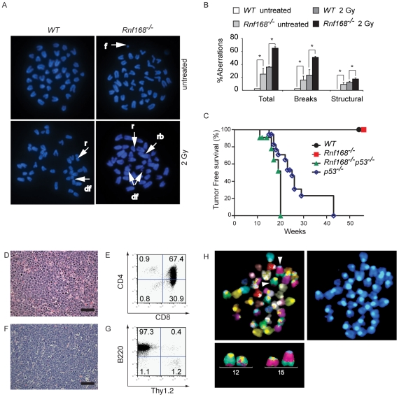
Figure 8. Rnf168 maintains genomic integrity and suppresses cancer.
(A and B) Metaphase spread analysis of Rnf168−/− and WT B-cells. Representative data (A) and the percentage of aberrations (B) are shown. Three independent experiments were performed. A minimum of 40 metaphase spreads of untreated or irradiated Rnf168−/− and WT cells were analyzed. *p<0.05. f = acentric fragment, r = ring, rb = Robertsonian translocation, df = double acentric fragment. (C) Kaplan Meier tumor-free survival analysis for cohorts of WT (n = 56), Rnf168−/− (n = 50), p53−/− (n = 18) and Rnf168−/−p53−/− (n = 11) mice. A statistically significant difference was observed between the tumor-free survival of Rnf168−/−p53−/− and p53−/− mice (p = 0.0096, log-rank test). (D and E) H&E staining (D) and FACS analysis (E) of a thymoma from an Rnf168−/−p53−/− mouse. (F and G) H&E staining (F) and FACS analysis (G) of a B-cell lymphoma (B220+) from an Rnf168−/−p53−/− mouse. (H) Chromosomal translocations observed in an Rnf168−/−p53−/− lymphoma. Clonal reciprocal chromosomal translocations t(12;15) and t(15;12) are shown. Scale Bars: 50 µm; (D and F).

