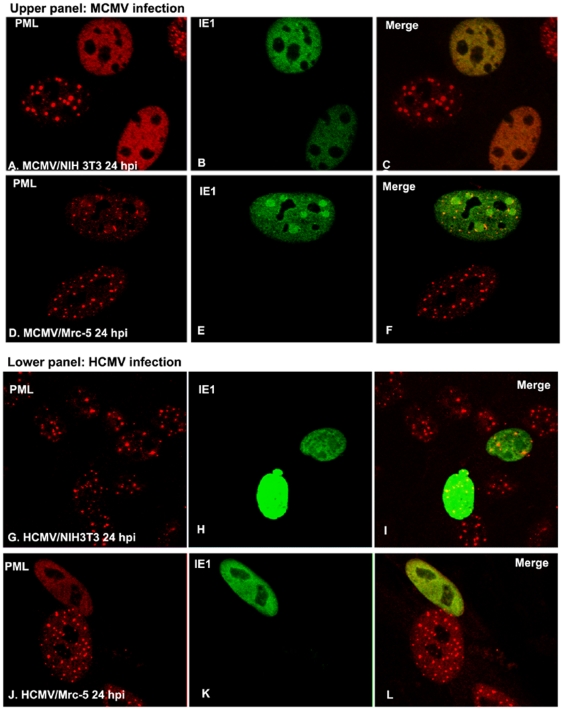
Figure 2. Immunofluorescent assay to show cytomegalovirus infection and ND10.
A–C: After MCMV infection in NIH3T3 cells for 24 hours, cells were stained with anti-PML antibody (rabbit) to show ND10 (in red) (A); anti-IE1 antibody (mouse) was used to show IE1 (in green) (B); the merged picture is shown in C. D–F: After MCMV infection in Mrc-5 cells for 24 hours, cells were stained with anti-PML antibody (rabbit) to show ND10 (in red) (D); anti-IE1 antibody (mouse) was used to show IE1 (in green) (E); the merged picture is shown in F. G–H: After HCMV infection in NIH3T3 cells for 24 hours, cells were stained with anti-PML antibody (rabbit) to show ND10 (in red) (G); anti-IE1 antibody (mouse) was used to show IE1 (in green) (H); the merged picture was shown in I. J–L: After HCMV infection in Mrc-5 cells for 24 hours, cells were stained with anti-PML antibody (rabbit) to show ND10 (in red) (J); anti-IE1 antibody (mouse) was used to show IE1 (in green) (K); the merged picture is shown in L.

