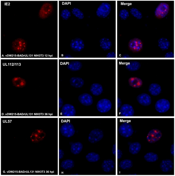
Figure 5. Immunofluorescent assay to detect HCMV proteins after infection in mouse cells.
A–C: Detection of HCMV IE2 in red (A), DAPI to show total cells (B), and the two merged in C. D–F: Detection of UL112/113 in red (D), DAPI to show total cells (E), and the two merged in F. G–I: Detection of UL58 in red (G), DAPI to show total cells (H), and the two merged in I.

