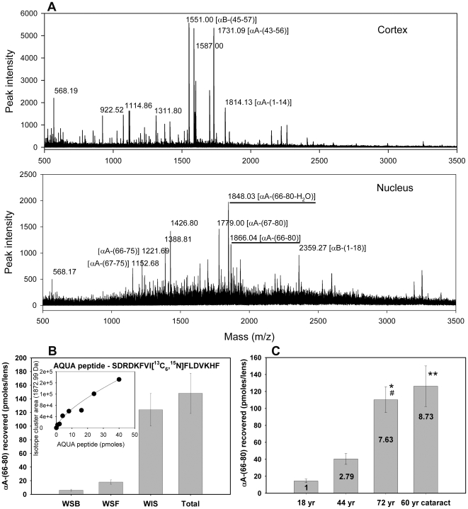
Figure 1. Distribution of the αA-(66-80) peptide in the lens.
(A) MALDI qTOF MS/MS analysis of LMW peptides isolated from the cortex and nucleus of a >70-year-old human lens as described under methods. The αA-(66-80) peptide characterized in this study is highlighted in the panel labeled – nucleus. (B) αA-(66-80) peptide concentrations in a >70-year-old lens fractions (WSB, water-soluble bound; WSF, water-soluble free; WIS, water-insoluble sonicated fractions). The amount of the peptide was estimated using 13C 15N-labelled spiked synthetic peptide standards (AQUA peptide) during peptide isolation. The inset figure is the standard curve for the isotope cluster area of the AQUA peptide used to determine in vivo peptide concentration. (C) Concentrations of αA-(66-80) peptide in human lenses of 18-, 44-, 60- and 72-year age groups. Lenses that were 60 years old were cataract lenses. Values within the bar show the multiple increased concentrations as compared to the 18-year-old lens. The values are mean ± SD of three analyses. * P<0.001 compared to 18 year, # P<0.01 compared to 44 year, ** P<0.001 compared to 18 and 44 year. P value determined using ANOVA. These results show an age-dependent increase in the concentration of αA-(66-80) peptide in human lenses and these peptides are associated with the WIS fraction. The cataract lenses show a higher level of αA-(66-80) peptide than the expected level of the peptide in age-matched non-cataract lenses.

