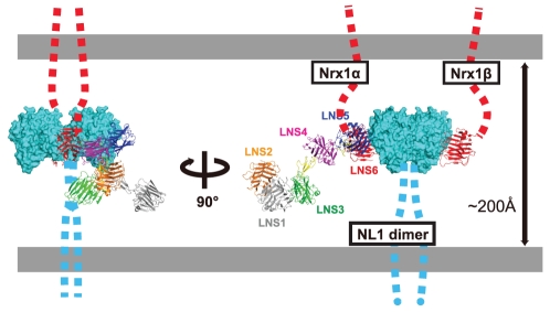
Figure 8. Schematic rendering of the Nrx1α/NL1 complex in the synaptic cleft.
Hypothetical NL1 dimer simultaneously bound by Nrx1β and Nrx1α from opposite sides are depicted in two different views. For Nrx1α-NL1 association, structural models of the NX1α(III)/NL1 complex (Fig. 6B) and the NX1αEC (Fig. 7E) are combined. In this rendering, LNS1+EGF1 segment of the Nrx1α was modeled but the position was determined arbitrary. The linker segments connecting toward cell membrane in both proteins are represented by dotted lines. The Nrx1/NL1 complex structure is drawn roughly in scale to the length of the synaptic cleft (∼200 Å).

