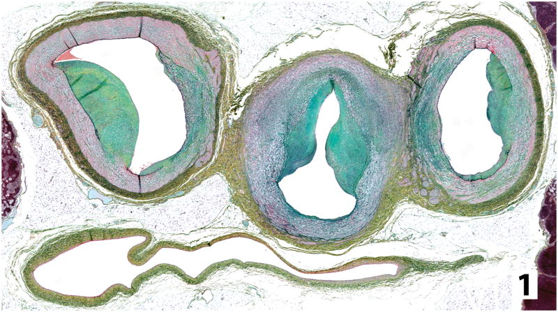
Figure 1.
Transverse histologic section from an untreated dog with MPS-I through distal aorta (central) just beyond branch points of external iliac arteries (left and right). Asymmetric areas of intimal sclerosis containing GAG (blue-green) infringe on vessel lumens and the normal smooth muscle (red) of the vessel is disrupted. Vena cava is below the arteries (Movat’s modified pentachrome).

