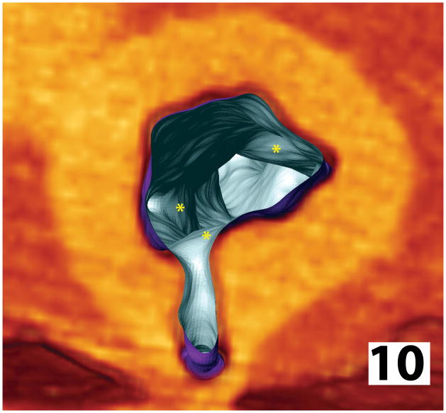
Figure 10.
Reconstruction of sublumbar aortic microCT scan from a non-tolerant dog with MPS-I that had received low doses of IdU. Areas of intimal plaque formation (asterisks) are localized just beyond arterial branch points and taper distally along the aortic wall, with some thickening at the os of these branches and luminal narrowing. In contrast, vessels from normal animals are cylindrical structures with smooth interior surfaces, constant luminal diameters and have a uniform circumferential wall thickness.

