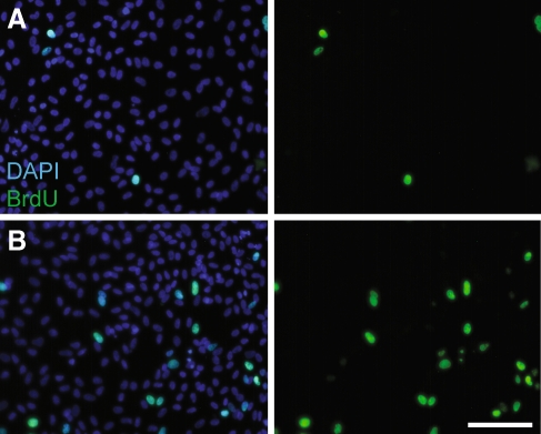
FIG. 1.
Decreased proliferation in epithelial cultures after treatment with VPA. Cultures of dissociated utricular supporting cells were treated for 24 h with 1 mM VPA and proliferating cells were labeled by the addition of BrdU for the final 4 h in vitro. Cultures were stained with DAPI (blue, left) and immunolabeled for BrdU (green, left and right). Shown are representative images of A VPA-treated and B control cultures. Scale bar 100 μm.

