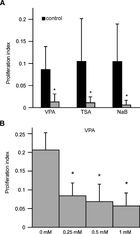
FIG. 2.
Quantification of proliferation in epithelial cultures following treatment with HDAC inhibitors. Cultures were treated for 24 h with VPA (1 mM), TSA (100 nM), or NaB (1 mM). Following fixation and histological processing, proliferation indices (BrdU-labeled cells/total cells per 10,000 μm2) were quantified for randomly selected image fields. A Cultures treated for 24 h with all HDAC inhibitors displayed >80% reduction in proliferation. B Dose–response relationship for treatment with VPA. Dissociated cultures were treated at varying doses (0, 0.25, 0.5, and 1 mM) for 24 h, and BrdU was added for the final 4 h in vitro. Increasing concentrations of VPA caused dose-dependent decreases in the proliferation index. Error bars: standard deviation, *p < 0.001.

