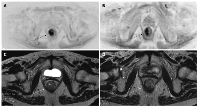
Figure 2.
Diffuse weighted imaging and cancer (arrows) of the lower rectum. At initial MR staging T3N1. After neoadjuvant therapy, MR restage was T3N0. At pathology: T3N1 (only one small metastatic mesorectal lymph node). Diffusion weighted imaging (DWI) (A) and axial T2w (B) images show the tumor (arrows) before chemoradiotherapy. The DWI image allows for better recognition of the lesion. DWI (C) and axial T2w (D) images after chemoradiotherapy show a reduction in the dimensions of the lesion (arrows).

