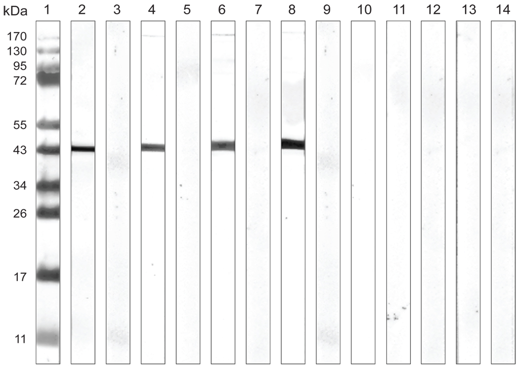
Figure 3. Western blot analysis of plasma samples.
C. trachomatis EBs of the human serovar F were used as the antigen. Lane 1) Molecular weight standards; Lane 2) control mAb E-4 to the C. trachomatis MOMP; Lanes 3), 5), 7), 9), 11), and 13) are plasma samples from animals Mmu-0.2, Mmu-0.9, Mmu-3.9, Mmu-2.5, Mmu-4.8 and Mmu-5.3 before immunization, respectively; Lane 4), 6), 8), 10), 12), and 14) are plasma samples from animals Mmu-0.2, Mmu-0.9, Mmu-3.9, Mmu-2.5, Mmu-4.8 and Mmu-5.3 two weeks after the third immunization, respectively.

