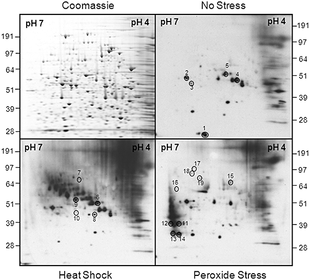
Fig. 8.

Identification of ubiquitinated proteins in C. albicans using a proteomic screen. C. albicans THE1 cells were grown for 5 h to mid-exponential phase and then exposed to stress for 1 h. Protein extracts were run on replicate 2-D gels, which were either stained with Coomassie blue or subjected to Western blotting with an α-ubiquitin antibody: Western blots of no stress control; peroxide-treated cells (50 mM H2O2); heat-shocked cells (30–42°C). Autoradiographs were aligned with the Coomassie-stained gels, spots chosen for analysis, and the corresponding proteins identified by tryptic digestion and LC-MS/MS (see text). The sample reference numbers for ubiquitination targets are shown (Table 2).
