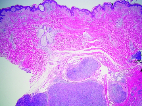
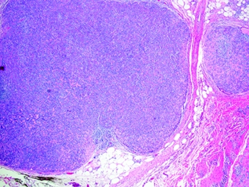
Figures 3A and 3B.
A) Overlying normal epidermis with underlying multiple, well-circumscribed, dermal nodules consisting of basophilic cells (hematoxylin-eosin [H&E], magnification 20X). B) Increased powered view of a dermal nodule with cords of basaloid cells arranged in a trabecular pattern with eosinophilic fibrous strands and numerous lymphocytes (H&E, magnification 40x).


