
FIGURE 5:
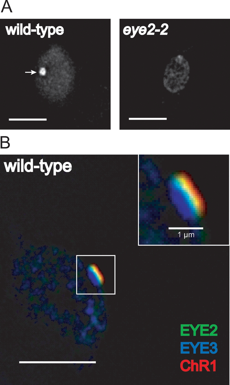
EYE2 localizes to a region corresponding to the chloroplast envelope in the eyespot. (A) Immunofluorescence micrographs of wild-type (left) and eye2–2 cells (right) stained with antibody directed against EYE2. EYE2 localizes to the eyespot (arrow) in wild-type cells, whereas staining is absent in the insertion mutant. (B) Combined immunofluorescence micrograph of wild-type C. reinhardtii cell stained with antibodies against EYE3 (blue), EYE2 (green), and ChR1 (red). Inset shows a close-up view of the eyespot area. EYE2 staining appears between the pigment granule layers and the plasma membrane–localized photoreceptor patch. Bars, 5 μm unless otherwise noted.