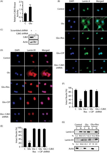
FIGURE 2:
Glutamate stimulation triggers dispersion of lamin A and lamin B1 in a Cdk5-dependent manner in HT22 cells. (A) Glutamate stimulates Cdk5 kinase activity. Cdk5 was immunoprecipitated from either control (column 1) or glutamate-treated HT22 cells (for 30 min), and the kinase assay was conducted using Cdk5 substrate peptide as described in Materials and Methods. C, control; Glu, glutamate. (B and D) HT22 cells were treated with 5 mM glutamate for 4 h, followed by immunostaining with lamin A (B, green) or lamin B1 (D, red) and DAPI (blue) as described in Materials and Methods. Roscovitine (10 μM) or TAT-CIP (200 nM) was added 0.5 h before glutamate treatment. Cdk5 shRNA lentivirus was added to infect cells for 30 h, followed by glutamate treatment. Representative pictures are shown. Scale bar, 20 μm. (C) Cdk5 levels in HT22 cells on lentiviral infection of Cdk5 shRNA for 30 h. (E) Percentage of cells showing lamin A dispersion was counted as described in Materials and Methods. (F) Percentage of cells showing lamin B1 dispersion was counted. Bar graphs shown are mean ± SD (*p < 0.05, *p < 0.01, ***p < 0.001). (G) Subcellular fractionation of lamin A and lamin B1 in glutamate-treated HT22 cells in the absence or presence of 10 μM roscovitine as described in Materials and Methods. The percentages of nuclear and cytoplasmic lamins were calculated for each sample as shown. Actin is the cytoplasmic marker. N, nuclear fraction; C, cytoplasmic fraction.

