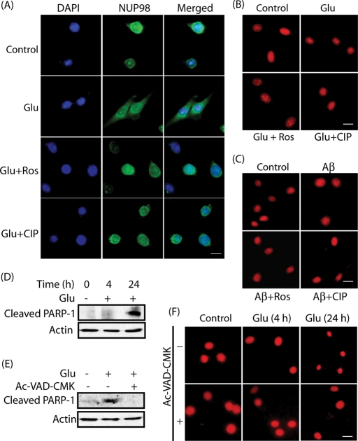
FIGURE 6:
Lamin dispersion precedes apoptosis in glutamate-treated HT22 cells. (A) HT22 cells were treated with 5 mM glutamate for 4 h in the absence and the presence of 10 μM roscovitine or 200 nM TAT-CIP, followed by immunostaining with NUP98 (green) and DAPI (blue). Representative pictures are shown. Scale bar, 20 μm. (B) HT22 cells were treated with 5 mM glutamate for 4 h in the absence and the presence of 10 μM roscovitine or 200 nM TAT-CIP, followed by staining with PI (red). (C) Cells treated with 25 μM Aβ25–35 were stained with PI (red). (D) Cleaved PARP-1 levels in glutamate-treated HT22 cells (4 h and 24 h). (E) Cleaved PARP-1 levels in glutamate-treated HT22 cells in the absence and presence of 40 μM Ac-VAD-CMK. (F) HT22 cells were treated with 5 mM glutamate for 4 h or 24 h in the absence and the presence of 40 μM Ac-VAD-CMK, followed by PI staining (red).

