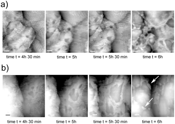
Figure 6. AFM images of H. walsbyi external capsule disruption during the drying process.
Height images are enlargements of two representative regions (white boxes in Fig. 5b time t = 4 h 30 min) showing capsule evolution during the drying process. The capsule features holes or lacerations that break up over time, gradually increasing in size. At the end of the process remnants of the capsule can still be seen, mainly located in the valleys between the protrusions (white arrows time t = 6 h). Scale bar 100 nm.

