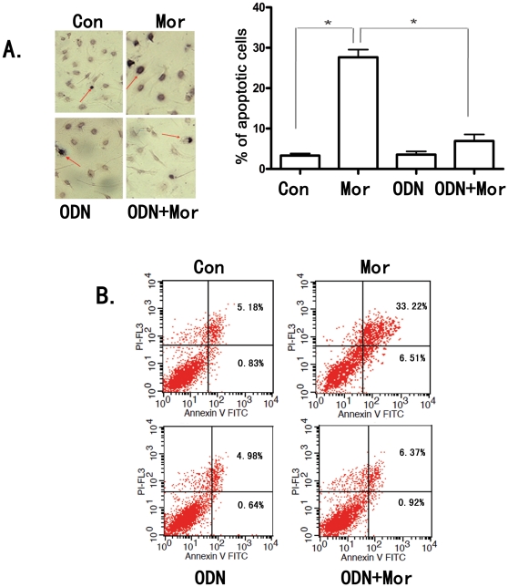
Figure 3. Inhibtion of TLR9 by CpGODN2088 blocked morphine-induced apoptosis in wild type microglia.
WT microglial cells were exposed to 5 µM CpGODN2088 (ODN) for 1 hr and then treated with morphine at 10 µM for 24 hr. (A). Apoptotic cells were determined by TUNEL assay as in Fig. 2. Representative light microscopic images showed TUNEL-positive microglia (red arrow head) Results represent mean ± SD of three independent experiments. * p<0.01 compared with indicated groups. (B). Cell apoptosis was also assayed by flow cytometry after staining with annexin V and propidium iodide as described under “Materials and Methods”. These results are representative of three independent experiments.

