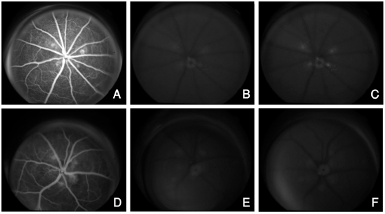
Figure 1. Bioimaging with NP-angiography showing GFP expression using the Topcon camera with fluorescein angiography filter settings.
Late phase FAs (A and D) show the CNV lesions prior to injection of NP. Autofluorescent images taken prior to injection of NP reveal minimal background fluorescence of the CNV lesions (B and E). Injection of targeted NP carrying a GFP plasmid (NP-GFPg) causes increased fluorescence of the CNV lesions from GFP expression (C) whereas non-targeted NP carrying a GFP plasmid (ntNP-GFPg) does not cause any increase in the intensity of fluorescence of the CNV over background autofluorescence (F).

