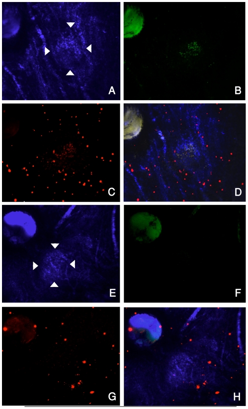
Figure 2. Choroidal flatmounts showing accumulation of rhodamine labeled NP and expression of GFP plasmid in the CNV.
The CNV lesions are delineated by arrowheads in bright field images with false blue color (A and E). FITC-filtered images highlight the GFP expression one day after systemic injection of Rd-NP-GFPg (B) whereas non-targeted NP (Rd-ntNP-GFPg) does not induce GFP expression in CNV (F). Cy3-filtered images highlight that rhodamine-labeled NP (Rd-NP-GFPg) accumulates in the CNV (C), while rhodamine-labeled non-targeted NP (Rd-ntNP-GFPg) does not (G). Some particles can be visualized circulating in the choroidal vessels. Overlay of images A–C is presented in panel D and overlay of E–G is shown in H.

