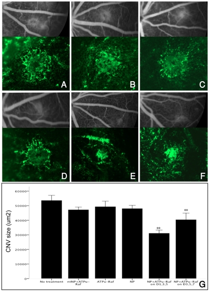
Figure 3. Late phase fluorescein angiography (FA) and choroidal flatmounts (x10) two weeks after laser photocoagulation.
Representative lesions are from the control group (A–D) and the NP-ATPμ-Raf treated group (E and F). Group (A) received no treatment; (B) received intravenous injection of non-targeted NP containing ATPμ-Raf on days 1, 3, and 5 after laser CNV creation; (C) received intravenous injection of ανβ3 targeted-NP without ATPμ-Raf gene on days 1,3, and 5; (D) received injection of ATPμ-Raf gene without NP on days 1, 3, and 5; (E) received injection of ανβ3 targeted-NP containing ATPμ-Raf (NP-ATPμ-Raf) on days 1, 3, and 5; and (F) received injection of NP-ATPμ-Raf on days 3, 5, and 7. NP-ATPμ-Raf treated groups (E and F) had significantly lower grade CNV lesions on FA grading and smaller CNV size compared to the control group (A–D). No statistically significant difference in size was noted between the control groups A–D. Quantification of the CNV size on choroidal flat mounts is shown in (G). *P<0.01. Data are expressed as the mean ± SE.

