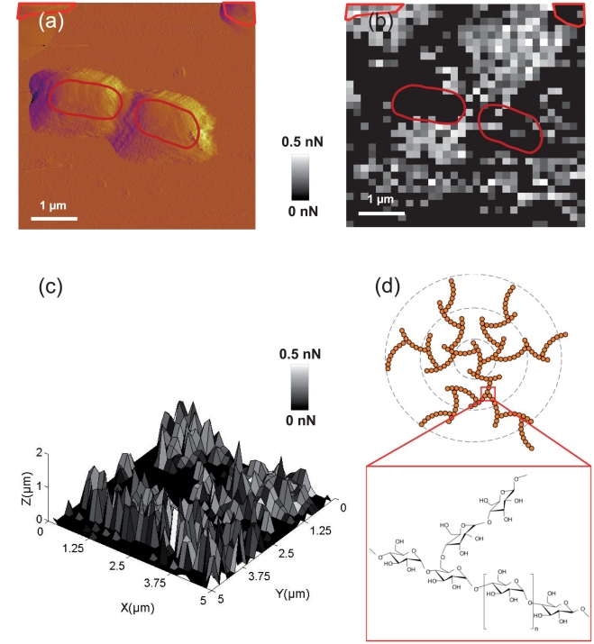
Figure 7. Maps of adhesive properties of P. fluorescens, and glycogene structure.
(a) Deflection image of Pseudomonas fluorescens taken before FVI experiment, the red zones correspond to bacterial cells location. (b) Adhesion force map (F-range = 0–0.5 nN) for the last adhesion force rupture measured on bacteria located on the surface as indicated in panel a. (c) Three dimensional map of adhesive properties of P. fluorescens combining the last adhesion force and last rupture distance corresponding to uncoiled exopolymers. (d) Fractal structure of the poly glycogene and chemical formula.

