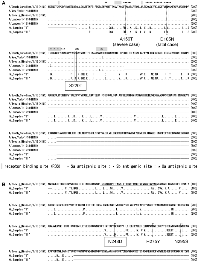
Figure 4. HA and NA sequence alignments between the 1918 H1N1 and 2009 pdm H1N1 viruses.
(A) The HA alignment between the consensus sequences obtained for samples “I” and “II” and the five available sequences of 1918 H1N1 viruses. Also represented on this alignment are the major substitution (S220T) between samples “I” and “II”; A156T and D185N observed in fatal or severe cases; the antigenic sites Sa (*), Sb (▴), and Ca ( ); the receptor binding site (highlighted in grey); and the cleavage site (underlined). (B) The NA alignment between the consensus sequences obtained for samples “I” and “II” and that of the 1918 A/Brevig_Mission/1/18 (H1N1) virus. Also represented on this alignment are the major substitution (N248D) between samples “I” and “II,” H275Y and N295S causing oseltamivir resistance, and the stalk region (underlined).

