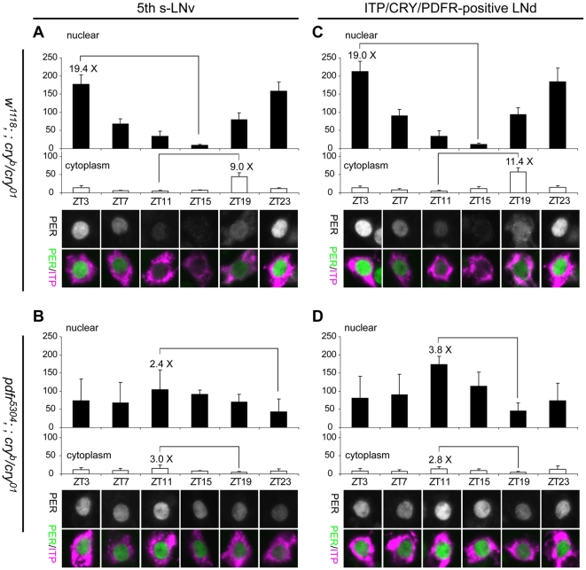
Figure 6. LD Molecular rhythms in the 5th s-LNv and LNd are deranged in the double mutants.
At various time-points, PER levels were monitored in the nucleus (filled histograms) and cytoplasm (open histograms) of the 5th s-LNv (A, B) and the ITP(+) LNd (C, D). (A) In the 5th s-LNv of w1118; ; cryb/01, PER levels in the nucleus and cytoplasm are robustly cycling: nuclear amplitude rhythm – 19.4-fold; cytoplasmic amplitude rhythm – 9.0-fold. ANOVA test revealed that the differences in nuclear staining levels are significant (P<0.0001). (B) In the 5th s-LNv of pdfr5304; ; cryb/01, PER staining is always found in the nucleus with very low amplitude rhythms and no phase difference between nucleus and cytoplasm: nuclear amplitude rhythm –2.4-fold; cytoplasm amplitude rhythm – 3.0-fold. ANOVA test revealed that the difference in this group is significant (P = 0.03). (C) In the ITP(+) LNd of w1118; ; cryb/01, PER levels in the nucleus and cytoplasm are robustly cycling, nuclear amplitude rhythm – 19.0-fold; cytoplasmic amplitude rhythm – 11.4-fold. ANOVA test revealed that the difference in this group is significant (P<0.0001). (D) In the ITP(+) LNd of pdfr5304; ; cryb/01, PER staining is always found in the nucleus with very low amplitude rhythms and no phase difference between nucleus and cytoplasm: nuclear amplitude rhythm – 3.8-fold; cytoplasmic amplitude rhythm – 2.8-fold. ANOVA test revealed that the difference in this group is significant (P<0.0001). Results from post-hoc statistical tests are presented in Table 2.

