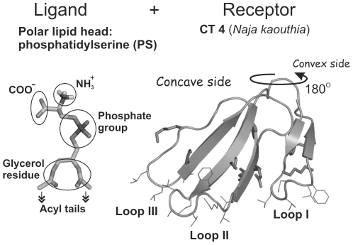
Figure 1. Three-dimensional structures used in molecular docking.
Spatial model of PS head group (hPS) with two-carbon acyl moieties in place of the full-length acyl chains (“ligand”) is displayed on the left panel. X-ray structure of CT A3 from Naja atra (complete sequence analogue of CT 4 from Naja kaouthia) (“receptor”, right panel) is drawn in stick mode. β-Strands of CT are indicated with arrows. Side chains of the membrane-binding hydrophobic residues, along with the charged ones surrounding loops I-III, are shown. Functional groups of hPS are indicated.

