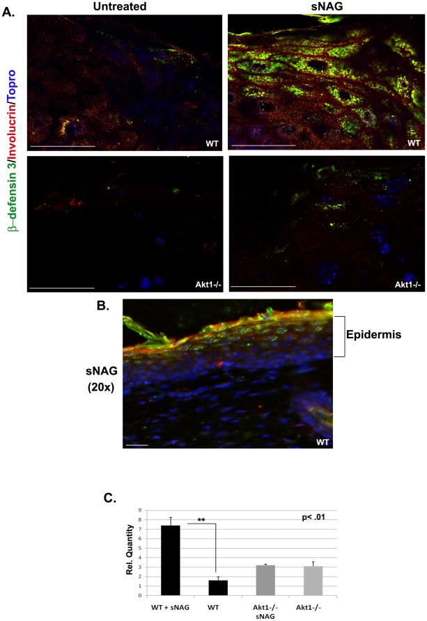
Figure 3. sNAG induced defensin expression in vivo requires Akt1.
(A) Paraffin embedded sections of cutaneous wounds harvested on day 3 post wounding from both WT (n = 3) and Akt1 mice. Wounds were either untreated or treated with sNAG membrane. Immunofluorescence was performed using antibodies directed against β-defensin 3 (green), Involucrin (Red), and Topro (Blue). (B) Paraffin embedded section from WT treated with sNAG harvested on day 3. Immunofluorescence was performed using antibodies directed against β-defensin 3 (green), Involucrin (Red), and Topro (Blue). This lower magnification (20×) is included to better illustrate the epidermal layers expressing β-defensin 3. Scale bars = 50 µm. (C) Quantitation of β-defensin 3 expression from paraffin embedded sections was performed using NIH ImageJ software. Experiments were repeated three independent times and p values are shown.

