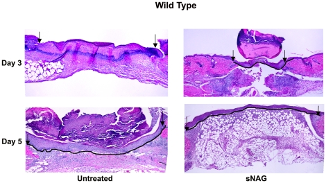
Figure 4. sNAG treatment increases wound closure in wild type mice (A).
H&E staining of wound tissue sections derived from C57Bl6 wild type animals either untreated or treated with sNAG membrane. The day post-wound is indicated to the left of each panel. The solid black line follows the keratinocyte cell layer indicating wound closure. Black arrows indicate the margin of the wound bed.

