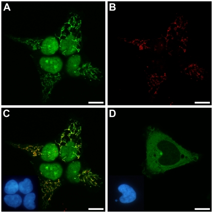
Figure 1. Subcellular localization of EGFP-tagged RNase ZL and RNase ZS in 293 cells.
(A) RNase ZL-EGFP; (B) immunofluorescence localization of subunit I of cytochrome c oxidase (mitochondrial staining) in the cells shown in (A); (C) overlay of (A) and (B) with nuclei shown as inset; (D) RNase ZS-EGFP with nuclei shown as inset. Confocal laser scanning microscopy (scale bar, 10 µm); insets showing nuclei by epifluorescent microscopy.

