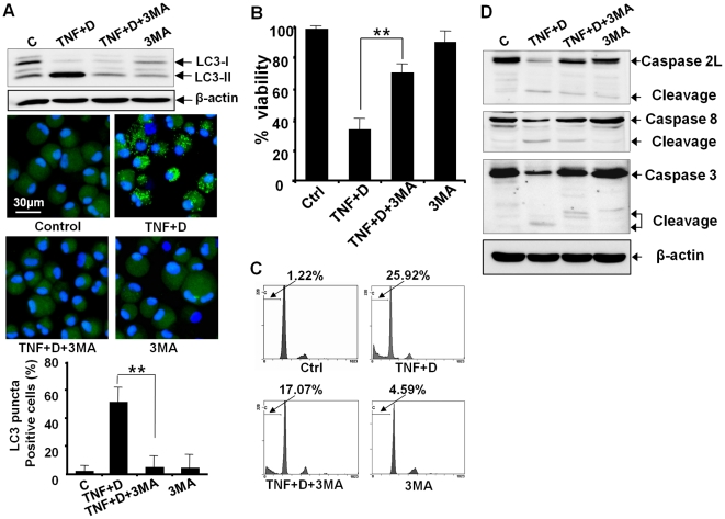
Figure 6. Inhibition of autophagy by 3MA protects chondrocytes against apoptosis as well as autophagy.
Cells were pretreated with 3MA for 24 h, and were further exposed to TNF-α in the presence of the CK2 inhibitor DRB for 24 h. 3MA, 3-methyladenine. (A) A western blot assay showed that 3MA suppressed the conversion of LC3 from LC-3I to LC-3II. Confocal microscopy also demonstrated that 3MA suppressed the appearance of a punctuate LC3 pattern. The graph showing the quantification data of the fraction of cells with puncta supported that 3MA significantly suppressed puncta formation (** P<0.01). β-actin, a loading control. (B) A viability assay showed that 3MA significantly protected chondrocytes against cell death (** P<0.01). (C) Flow cytometry demonstrated that 3MA prevented the accumulation of subdiploid cells. (D) A western blot assay showed that 3MA protected chondrocytes against the activation of caspase-2L, -8, and -3. See Figure 2 for other definitions.

