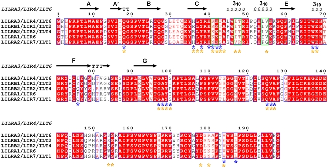
Figure 1. Multiple amino acid sequence alignment of the D1 and D2 domains of Group 1 LILRs.
The secondary structure elements of LILRA3 D1 are shown above the sequences. The asterisks indicate the residues involved in the HLAIs binding of LILRB1 and LILRB2 (blue for LILRB1 and yellow for LILRB2). The residues previously hypothesized to be critical to form the 310 helices region in domain 1 of Group 1 LILRs are demarcated with green.

