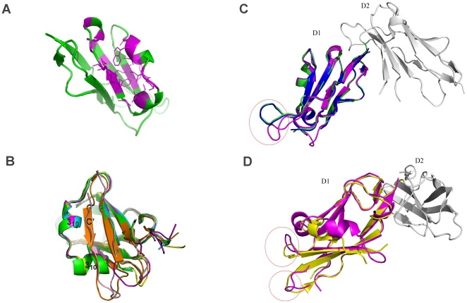
Figure 4. Hydrophobic pocket of LILRA3 D1 and comparison of the crystal structures of LILRA3 D1, LILRB1 D1, and LILRB2 D1.
A. The hydrophobic pocket of LILRA3 D1. The hydrophobic residues located around this area are colored with magenta. B. The helix-strand transition between LILRB1/LILRB2/LILRA3 and LILRA2/LILRA5: magenta, LILRA3; cyan, 1GOX (LILRB1); green, 1UFU (LILRB1); yellow, LILRB2 in complex with HLA-G1 (2DYP); orange, 2D3V (LILRA5); and light pink, LILRA2 C. The difference in the loop region of the LILRA3 D1 (pink) superposed on LILRB1 D1D2 (1GOX-green and 1UFU-blue). D. The difference of the loop region of LILRA3 D1 (pink) superposed on the LILRB2 D1D2 (2GW5-yellow).

