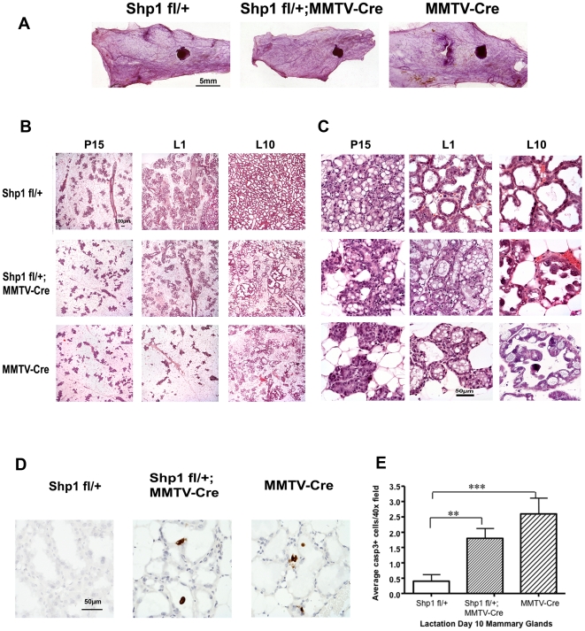
Figure 2. Impaired differentiation and precocious involution of mammary epithelial cells in mice expressing the MMTV-Cre transgene.
(A) The number 4 mammary glands from Shp1 fl/+ (control), Shp1 fl/+/;MMTV-Cre, and MMTV-Cre mice were isolated from 10-week old virgin mice and subjected to whole mount analysis as described in the Materials and Methods. Scale bar = 5 mm. (B, C) Six-week old Shp1 fl/+ (control), Shp1 fl/+;MMTV-Cre, and MMTV-Cre female mice were mated with male wild type mice. The number 4 mammary glands were removed at pregnancy day 15 (P15), lactation day 1 (L1), and day 10 (L10), and processed for histological analysis as described in the Materials and Methods. Images were taken under lower (B) and higher (C) magnification respectively. Scale bars represent 100 µm in (B) and 50 µm in (C). Similar results were seen from at least two different mice for each genotype. (D, E) Analysis of apoptotic cells in L10 mammary glands as described in (B, C). (D) Sections of L10 mammary glands from indicated mice were stained with antibody against cleaved caspase 3 (see Materials and Methods). Cells positive for cleaved caspase 3 are stained dark-brown. Images were taken using 40X objective lens. Scale bar = 50 µm. (E) Quantification of caspase 3 positive cells from (D) (see Materials and Methods). Two mice from each genotype were analyzed. **p<0.01 and ***p<0.001.

