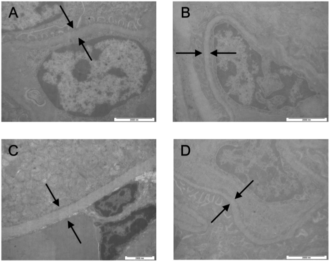
Figure 5. Ultrastructure of the glomerular basement membranes of the immunized animals after the Y. pestis challenge.
The control animals infected with Y. pestis were observed under a transmission electron microscope. Transmission electron micrograph of the glomerular basement membrane (GBM) of the control animal that that were only immunized with aluminum hydroxide adjuvant and then infected with virulent Y. pestis strain 141 (a), the animal immunized with EV76 (b), the animal immunized with subunit vaccine SV1 (c), and the animal immunized with subunit vaccine SV2 (d).

