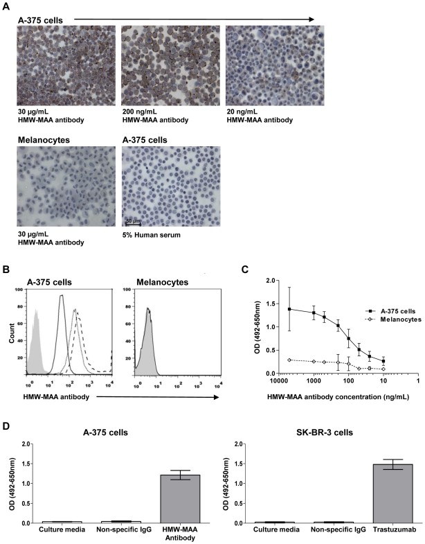
Figure 1. Development of a cell-based ELISA to detect tumor-specific antibodies.
(A) Immunocytochemistry of cytospin preparations demonstrates that an antibody against the melanoma-associated antigen HMW-MAA, expressed on the surface of A-375 metastatic melanoma cells, can detect the antigen at 30 µg/mL (top left), 200 ng/mL (top middle) and 20 ng/mL (top right), while no binding to melanocytes was seen (bottom left). IgG from 5% human serum (bottom right) did not bind to A-375 cells. (B) Flow cytometry analysis demonstrates specific binding of the HMW-MAA antibody to A-375 cells (left) at 30 µg/mL (dashed line), 200 ng/mL (dotted line) and 20 ng/mL (solid line), but not to melanocytes (30 µg/mL HMW-MAA antibody). Isotype control is shown in shaded grey histogram. (C) Detection of anti-HMW-MAA antibody binding to melanoma cells over a range of antibody concentrations compared to melanocytes by cell-based ELISA. (D) Melanoma-specific antibody HMW-MAA binding to A-375 cells compared to an IgG isotype control or to culture media (left) utilizing the cell-based ELISA. Breast cancer-specific antibody Trastuzumab binding to SK_BR-3 cells compared to an IgG isotype control or to culture media (right). Error bars in figures represent 95% confidence intervals.

