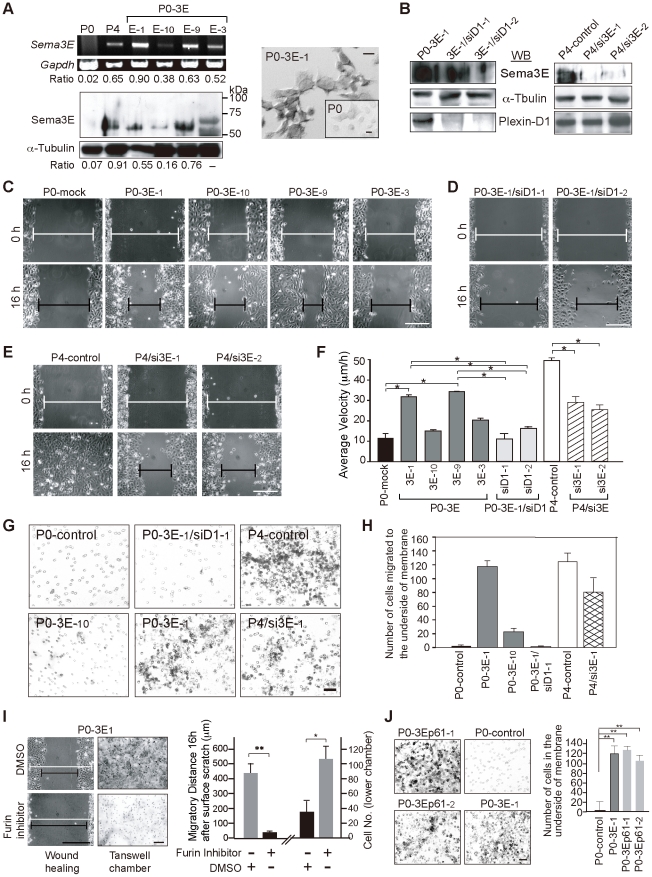
Figure 2. Sema3E mediates the invasive/migratory ability of OEC cells in vitro in a Sema3E-p61/Plexin-D1 dose-dependent manner.
A. Modified P0 cell lines (P0-3E-x) express variable levels of Sema3E as measured by semi-quantitative RT-PCR analysis (left top) and Western blotting (left bottom). Expression values were normalized to Gapdh and œ-Tubulin for mRNA and protein, respectively. RNA in situ hybridization probes by Sema3E in P0-3E-1 compared with P0 cells (right). B. Western blotting of Sema3E and Plexin-D1 in P4/si3E clones (RNAi-1, -2 for targeting two different sequences of Sema3E) and P0-3E-1/siD1 clones (RNAi-1, -2 for targeting two different sequences of Plexin-D1). C, D, E, F. Brightfield photomicrographs from wound-healing migration assays of various P0-3E (C), P0-3E-1/siD1 (D), and P4/si3E (E) cell lines at the beginning (0 h) and 16 hours after surface scratch. Wound edges were marked with white lines (0 h) or black lines (16 h). Scale bar, 0.5 mm. A summary of the average migrating velocity (µm/h) for each clone is plotted (F) with standard deviation (n≥12). *, P<0.05, paired t-test. G, H. Representative images (G) and graph summary (H) of the transwell chamber invasion assay (n≥6). Scale bar, 30 µm. I. The furin inhibitor, decanoyl-RVKR-chloromethylketone, alters migratory ability of P0-3E-1 cells in wound-healing and transwell chamber migration assays. DMSO was used as a control. White line, 0 h in wound-healing process; black line, 16 h after surface scratch. **, P<0.005, *, P<0.05, paired t-test, n = 5. Scale bar, 0.5 mm. J. Transwell chamber invasion assay for P0 cells stably expressing p61-Sema3E isoform (P0-3Ep61-1 and P0-3Ep61-2) compared with P0-3E-1 cells and P0 cells. **, P<0.005, paired t-test, n = 4. Scale bar, 0.3 mm.

