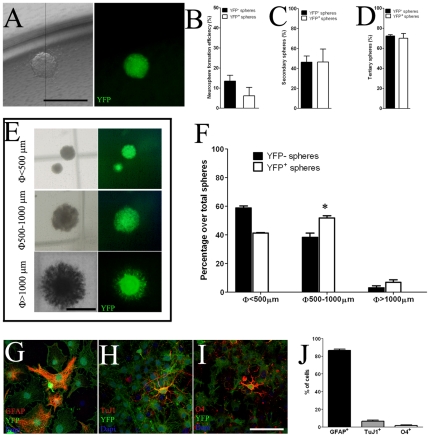
Figure 4. Embryonic AspM+ precursors give rise to aNSCs.
Panel A shows single neurospheres derived from YFP+ cells. Quantification of the clonal efficiency of YFP+ cells was obtained by plating single neural stem cells on 96 wells (n = 3 independent experiments). Percentages (± S.E.M.) of YFP− and YFP+ primary, secondary and tertiary neurospheres are plotted in panels B–D, respectively. Panel E shows representative images of YFP+ neurospheres of different sizes in the NCFC assay (<500 µm, 500–1000 µm and >1000 µm). AspM-CreERT2/Rosa26-YFP SVZ derived neural stem cells (n = 2 independent cell cultures) were propagated in vitro and then (after the 4th IVP) assayed by NCFC assay. The mean percentages (± S.E.M.) of both YFP− and YFP+ neurospheres of each size (n = 3 independent experiments for each cell culture) are plotted in panel F. Dissociated YFP+ neurospheres were capable to differentiate into astrocytes (G), neurons (H) and oligodendrocytes (I). Percentages (± S.D.) of each cell type are indicated in histogram on panel J. Scale bars: 1000 µm (panels A and E); 200 µm panels G–I. * <0.05, t-student.

