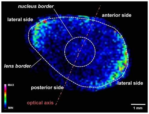
Figure 4. Molecular image of PC (34∶2) used to visualize structures in an ocular lens using matrix-assisted laser desorption ionization mass spectrometry imaging (MALDI MSI).
The lens cortex is the area between the nucleus and lens borders. Equatorial margins are clearly defined by the high abundance of phospholipids, and the lens nucleus is less visible in this case due to the changes in lipid composition in the lens.

