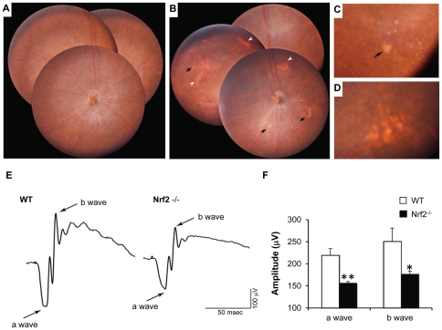
Figure 1. Early AMD-like degeneration in Nrf2−/− mice.
(A) Normal fundus photograph from a 12-month-old wild-type mouse. (B) Merged photos from central and peripheral retina from a 12-month-old knockout mouse, showing both dotted and patchy deposits (arrows) and RPE mottling (arrowheads). (C) Magnified picture showing spots with sharp outline and a large patchy deposit in the mid-peripheral retina (arrow). (D) Soft drusen-like deposits with larger size and ambiguous border. (E and F) Scotopic ERG recordings at +10 dB (25 cd·s/m2) flash intensity, showing significantly decreased a- and b-wave amplitudes in Nrf2 −/− mice when compared to age-matched wild-type mice (n = 6 per group; *P<0.05, ** P<0.01, Student's t-test).

