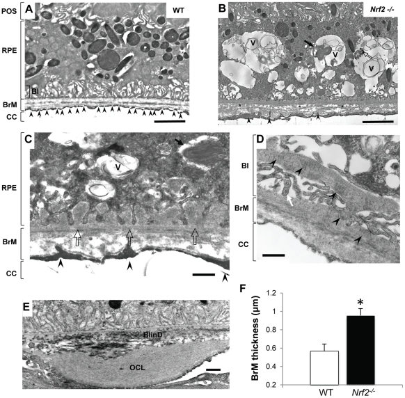
Figure 3. Ultrastructual changes in outer retina of aged Nrf2−/− mice.
(A) Electron micrograph of a 12-month-old wild-type mouse. Endothelial fenestrations of choriocapillaris (CC) were marked by arrowheads. (B) RPE of an Nrf2−/− mouse at 12-months showed large vacuoles (V) containing membranous debris, undigested POS (arrow) and melanin-containing materials (open arrow). BrM was thickened with disorganized collagen and elastin fibers (asterisks). (C) Basal infoldings were replaced by continuous basal deposits (open arrow). (D) Higher magnification showing basal laminar deposits and traverse-banded structure (arrowhead). (E) Extensively thickened outer collagenous layer (OCL) and basal linear deposits (BlinD). (F) Increased BrM thickness in aged Nrf2−/− mice. Data presented are average of measurements from 6 mice per group (mean ± SE) (* P<0.01, Student's t-test).

