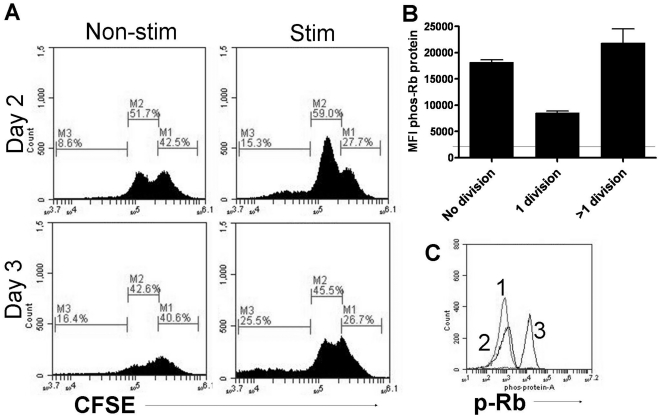
Figure 4. CDK4 activity in non-dividing cells.
A) Mixed myeloid cells expanded from bone marrow of C57Bl/6 mice were stained with CFSE prior to stimulation with the TLR7 agonist R848. After 48 and 72 hours cells were fixed and stained for phosphoryated Rb protein on the CDK4 specific site Ser 780. Cells in gate M1 are CFSE high and represent non-dividing cells. The percentage of cells within this gate remained approximately 26% for three days. Cells in gates M2 and M3 are CFSE low and represent cells that have gone through 1 or more than 1 division respectively. From day 2 to day 3 the % of M2 cells has decreased while the percent of cells in M3 has increased indicating ongoing proliferation. B) A histogram of MFI for phospho-Rb from each of the M gates indicates phosphorylation of Rb protein in all stages of division (line represents MFI of non-stimulated cells). C) Histogram of cells from M1 gate stained for phospho-Rb protein after 48 hours of R848 stimulation. Peak 1 is the isotype control of stimulated cells, peak 2 is the p-Rb staining of non-stimulated cells, and peak 3 is the p-Rb staining of R848 stimulated cells. These data are representative of two individual experiments.

