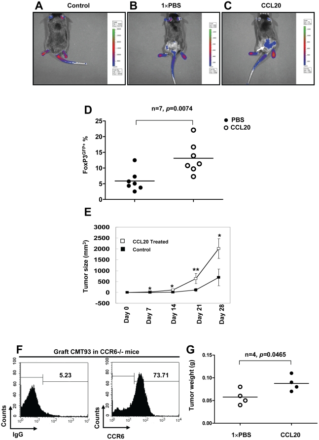
Figure 4. CCL20 recruited FoxP3+ Treg-cells to tumor mass and stimulated the growth of CRC in mice.
Mouse colorectal cell line, CMT 93 (1×10⁶) with or without 0.5 µg mouse recombinant mouse CCL20 in 100 µl PBS were injected s.c. into FoxP3GFP mice, and 0.5 µg recombinant mouse CCL20 in 100 µl PBS was injected s.c. weekly for 28 days. Control FoxP3GFP mice were only given 100 µl PBS injection. (A) Normal FoxP3GFP mouse was used as a technique control. (B, C) Green fluorescence of FoxP3GFP+ cells in mice treated PBS or recombinant mouse CCL20 (white arrow) were monitored using the IVIS Imaging System at day 28. (D) Numbers of tumor-infiltrating FoxP3GFP+ Treg-cells derived from grafted CRC in mice treated with PBS or recombinant mouse CCL20 were analyzed by Flow cytometry. (E) The sizes of tumors were measured with calipers at the indicated time points for 28 days. Injection of recombinant mouse CCL20 resulted in a significant increase in tumor sizes (n = 5). (F) Expression levels of CCR6 by grafted CRC in CCR6−/− mice. (G) CMT 93 (1×10⁶) with or without 0.5 µg mouse recombinant mouse CCL20 in 100 µl PBS were injected s.c. into CCR6−/− mice, and 0.5 µg recombinant mouse CCL20 in 100 µl PBS or 100 µl PBS was injected s.c. at every second day for 21 days. Tumor weight of CRC was measured at the end of experiment. Representative data are shown which had been reproduced in 3 independent experiments. * p<0.05, ** p<0.01, Student's t test.

